How ML Enhances Precision in Radiological Diagnostics
You might not realize just how much machine learning is reshaping the landscape of radiological diagnostics. By employing advanced algorithms, it's capable of identifying minute details in images that human eyes can easily miss. This not only boosts precision but also addresses the common pitfalls of human error. As ML continues to evolve, it raises questions about the future roles of radiologists and how these technologies will integrate into clinical workflows. What implications could this have for patient outcomes and the overall efficiency of healthcare?
If you're looking for assistance with this topic or have specific questions, feel free to reach out to us. We can connect you with experts who can provide tailored solutions and insights.
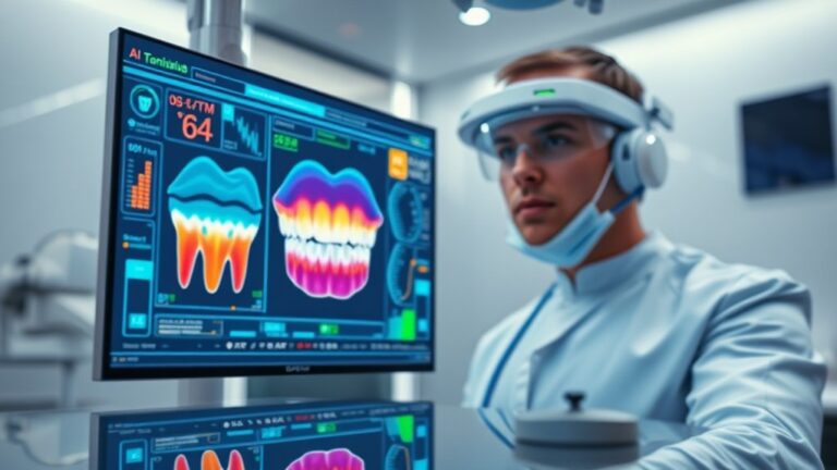
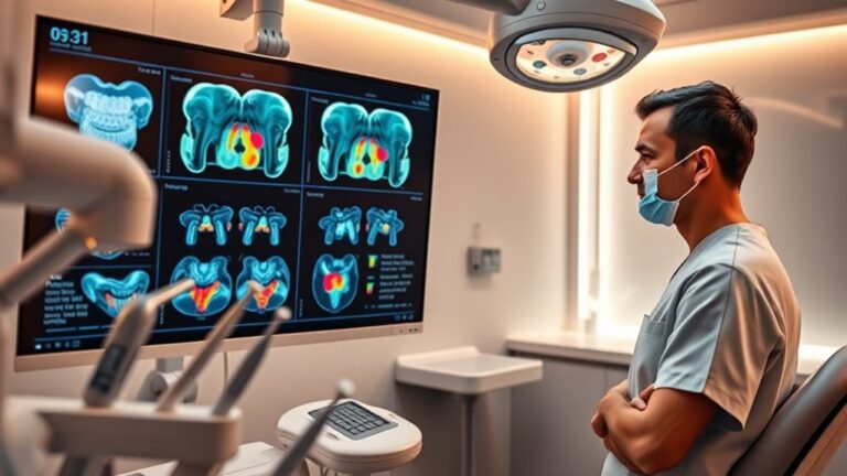
Machine Learning in Dental Radiology
In the domain of dental radiology, machine learning (ML) greatly enhances diagnostic capabilities by automating the detection of various dental pathologies. With accuracy rates reaching 91.5% in caries detection and 99.95% for periapical lesions, ML models notably improve diagnostic precision.
These automated pathology detection systems not only identify common issues but also accurately detect periodontal bone loss and maxillary sinusitis, ensuring thorough patient assessments. Furthermore, ML streamlines cephalometric analysis by automating the identification of key anatomical landmarks. This reduces manual labor and bolsters accuracy, allowing you to focus more on patient care.
With improved time efficiency, AI-driven systems accelerate the analysis of cephalometric radiographs, providing you with consistent and standardized results across multiple images. Additionally, ML's capability to achieve high accuracy in periapical lesion detection demonstrates its potential in transforming dental diagnostics.
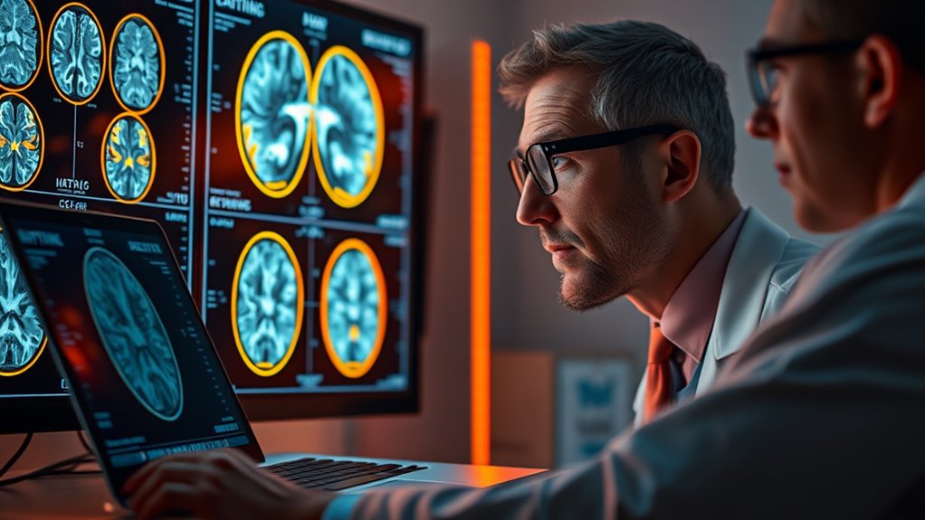
Enhancing Image Analysis Techniques
Leveraging advanced machine learning techniques notably enhances image analysis in radiology, allowing for more accurate and efficient diagnoses. Convolutional Neural Networks (CNNs) are essential in this process, employing CNN optimization strategies to improve image classification and detection. By recognizing complex patterns, these models can effectively distinguish between malignant and benign tissues, increasing the likelihood of early cancer detection. Moreover, the effectiveness of these models heavily relies on the quality of input data, underscoring the importance of high-quality datasets in achieving reliable outcomes.
Data augmentation techniques further strengthen the robustness of ML models. By artificially expanding datasets, you can expose your algorithms to a wider variety of conditions, reducing overfitting and enhancing generalizability. For instance, applying transformations like rotation and scaling to medical images guarantees that the model learns to identify abnormalities in diverse scenarios.
Additionally, advanced segmentation methods, such as U-Net and instance segmentation, enhance precision in identifying specific lesions. This capability is vital for tailoring treatment plans and improving surgical outcomes.
Reducing Human Error in Diagnostics
Reducing human error in diagnostics is essential for improving patient outcomes, and machine learning plays a pivotal role in this endeavor. By employing advanced algorithms, you can considerably reduce human oversight, which often stems from fatigue and the pressure of managing large volumes of cases.
Unlike humans, machine learning systems maintain consistent diagnostic accuracy, allowing for rapid processing of images without sacrificing performance. Machine learning excels at detecting subtle abnormalities that might elude even the most trained eyes, such as tumors in X-rays or lesions in MRIs. Its ability to analyze vast datasets allows it to identify complex patterns and correlations, resulting in more accurate diagnoses.
For instance, AI-assisted mammography screenings have demonstrated a 30% reduction in false positives while preserving high sensitivity for breast cancer detection.
Furthermore, machine learning algorithms enhance the overall diagnostic process by minimizing misdiagnoses and ensuring patients receive timely treatment. With predictive analytics, these tools can forecast disease progression and enable early interventions.
Improving Operational Efficiency
Machine learning not only enhances diagnostic accuracy but also greatly boosts operational efficiency in radiology departments. By leveraging AI algorithms, you can considerably enhance speed in image analysis. For instance, these algorithms can process and interpret an image every three seconds, a pace unattainable by humans. This rapid analysis is essential in emergency situations, where timely decisions can save lives. Furthermore, the use of deep learning techniques allows for even more sophisticated image recognition and interpretation.
Moreover, workflow automation plays an important role in optimizing radiological processes. AI streamlines nondiagnostic tasks like order entry and patient scheduling, freeing up valuable time for radiologists. Automated assignment of radiology protocols achieves high accuracy, allowing for more efficient workflow management.
Additionally, AI improves resource management by predicting workload and optimizing equipment utilization. This guarantees that resources are allocated effectively, especially during peak times. Reducing the need for repeat scans further optimizes operational efficiency, as AI minimizes misdiagnosis risks, ultimately saving time and healthcare costs.
Incorporating machine learning into your radiology department not only enhances the speed and accuracy of diagnostics but also guarantees that your team can focus on providing the best care possible.
Clinical Decision Support Advancements
Clinical decision support (CDS) tools increasingly play an essential role in enhancing the quality and appropriateness of radiological diagnostics. Mandated by the Protecting Access to Medicare Act (PAMA), these tools require physicians to consult appropriate use criteria (AUC), ensuring regulatory compliance that directly impacts reimbursement in radiology practices. This integration is particularly vital for advanced imaging orders such as CT and MRI, as it not only streamlines workflows but also aligns with MIPS standards.
AI integration further bolsters these CDS mechanisms by automating data analysis and reducing the likelihood of errors or misdiagnoses. For instance, studies show that implementing these tools can elevate the frequency of indicated studies from 64.5% to 82%, dramatically improving appropriateness scores for high-cost imaging. Furthermore, a recent study confirmed that CDS enhances imaging appropriateness by achieving a 3% increase in "usually appropriate" classifications. Additionally, the tools fit seamlessly into existing electronic health record systems, minimizing disruption while maximizing compliance.
Metrics of Accuracy and Precision
The evaluation of diagnostic accuracy and precision in radiology relies on a variety of metrics that illuminate the effectiveness of imaging techniques. Traditional metrics, such as sensitivity analysis and specificity evaluation, serve as foundational tools in gauging diagnostic performance. For instance, when diagnosing prostate cancer via MRI, you might observe sensitivity rates around 95% but specificity dipping to 50%. Confusion matrices further aid this evaluation, detailing true and false positives and negatives.
Additionally, incorporating information theory enhances these evaluations. Mutual information (MI) offers an innovative approach, modeling the diagnostic process as diagnostic channels that connect a patient's condition to the radiologist's interpretation. This method transcends biases inherent in traditional metrics, facilitating a clearer view of inter-reader agreement. The more information that flows through these channels, the more accurate your diagnosis becomes. Furthermore, information theory-derived measures provide alternatives to conventional statistics, enriching the analysis of diagnostic accuracy.
Moreover, metrics like ROC analysis, which evaluate varying cutoff points, complement these approaches by providing insight into error rates—estimated to be 3-5% in radiological diagnostics.
Understanding the interplay between these metrics not only elevates diagnostic accuracy but ultimately serves to improve patient care.
Frequently Asked Questions
How Does Machine Learning Handle Varying Quality in Medical Images?
You utilize image preprocessing techniques and quality assessment algorithms to effectively manage varying medical image quality. These tools enhance clarity, reduce noise, and guarantee accurate analysis, ultimately improving diagnostic outcomes and patient care.
What Types of Medical Images Benefit Most From ML Techniques?
Imagine unearthing hidden treasures in medical images! CT scans, MRI imaging, X-ray analysis, and ultrasound interpretation thrive with ML techniques like anomaly detection, image segmentation, and feature extraction for enhanced diagnostics and patient care.
Can ML Algorithms Be Trained on Small Datasets Effectively?
Yes, you can effectively train ML algorithms on small datasets by employing data augmentation techniques and transfer learning. These methods enhance model robustness and generalization, allowing you to maximize performance even with limited data availability.
How Do ML Models Ensure Patient Data Privacy and Security?
It's amusing, really—while data deidentification claims to protect privacy, consent complexities often obscure true understanding. You'll find that ML models utilize advanced techniques to guarantee patient data remains secure and confidential throughout the process.
What Are the Limitations of ML in Radiological Diagnostics?
You'll find that ML in radiological diagnostics faces limitations like algorithm generalizability issues and bias propagation, which can compromise accuracy. These factors lead to unreliable predictions, especially when applied to diverse, external datasets.
Conclusion
In summary, machine learning is revolutionizing radiological diagnostics, particularly in dentistry. With studies showing that CNNs can achieve up to 95% accuracy in detecting early caries, it's clear that these technologies not only enhance image analysis but also substantially reduce human error. By streamlining workflows and supporting clinical decisions, ML is not just a tool but a transformative force in radiology, making accurate diagnoses faster and more reliable than ever before.
If you're looking to navigate the complexities of incorporating machine learning into your practice, we encourage you to reach out to us for assistance. Getting expert help can save you valuable time, reduce stress, and ultimately improve your dental practice. Let us help you harness the power of these advanced technologies to enhance your diagnostic capabilities.