
Top 3D Imaging Solutions for Modern Orthodontic Practices
When you consider how practices using the iTero Digital Impression System have streamlined patient consultations, it's clear that 3D imaging is revolutionizing orthodontics. These technologies not only enhance diagnostic capabilities but also improve treatment planning and patient experiences. You might be wondering which specific solutions stand out in today's market and how they can impact your practice. Understanding their unique features could be the key to elevating your patient care and practice efficiency. If you need assistance navigating these options, feel free to contact us; we can connect you with experts who can provide tailored solutions for your practice.
Overview of 3D Imaging in Orthodontics
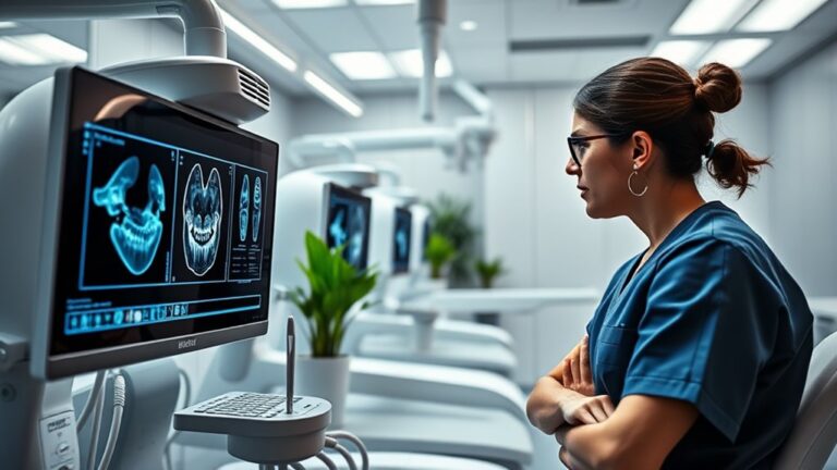
In orthodontics, the integration of 3D imaging technologies has revolutionized diagnostic and treatment processes, providing more than just a glimpse into the oral cavity.
By employing advanced 3D imaging applications like Cone Beam Computed Tomography (CBCT) and i-CAT FLEX, you can obtain high-definition visuals of teeth, bones, and soft tissues. This detailed imaging enhances your ability to diagnose issues such as impacted and supernumerary teeth, enabling a thorough examination of nerve and root health. Additionally, 3D imaging (CBCT) provides a comprehensive view of oral and maxillofacial structures, allowing for more accurate identification of complex dental anomalies.
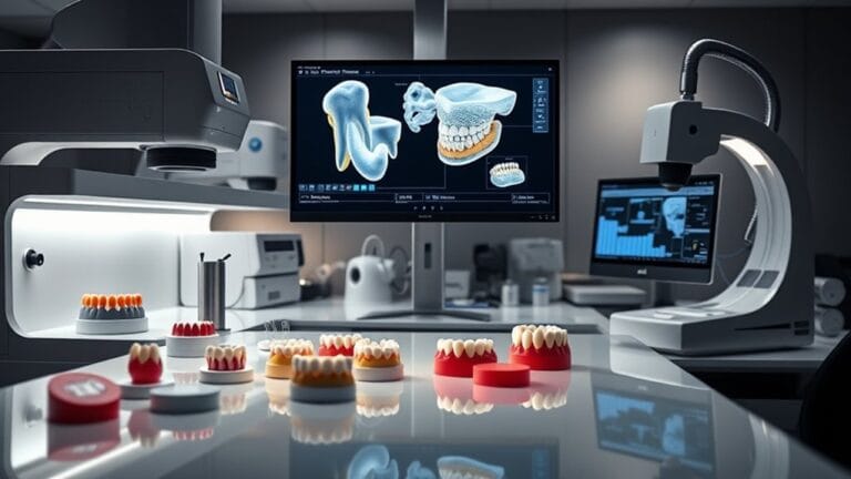
3D imaging is essential for refining the orthodontic workflow. It allows you to design custom orthodontic appliances tailored to each patient's unique anatomy.
With the ability to simulate treatment scenarios, you can predict outcomes more accurately, enhancing the quality of care you provide. Furthermore, regular scans help you track patient progress, facilitating timely interventions when necessary.
Benefits of 3D Dental Scanning
3D dental scanning offers significant advantages that enhance both diagnostic accuracy and treatment efficiency in orthodontics. With its ability to generate detailed, high-quality images of teeth and surrounding structures, you can diagnose issues like caries, gum disease, and fractures more effectively than with traditional X-rays. This technology facilitates thorough examinations, allowing you to spot problems that might otherwise go unnoticed.
In terms of treatment, 3D scanning streamlines office visits by reducing preparation time and enabling real-time simulations of various options. This not only shortens appointment durations but also enhances patient comfort, as they experience fewer invasive procedures. By using digital impressions, you can create high-quality prosthetics and accurately plan implant placements, which contributes to overall cost efficiency. Furthermore, the capability for early detection of dental issues ensures timely interventions, enhancing patient outcomes.
Additionally, 3D imaging promotes improved communication with dental specialists and laboratories. Sharing electronic files of digital impressions simplifies collaboration and enhances understanding of treatment plans through interactive displays.
Finally, the ability to monitor patient progress with easily retrievable scans guarantees that you can track outcomes effectively, reducing the likelihood of complications and additional treatments, ultimately leading to a more efficient practice.
Top 3D Imaging Technologies
Several cutting-edge 3D imaging technologies are revolutionizing orthodontics, enhancing diagnostic accuracy and treatment planning.
These advancements help you provide better care for your patients by enabling precise assessments and tailored treatment plans.
Here are three top technologies to evaluate:
- Cone Beam Computerized Tomography (CBCT): This technology uses a cone-shaped beam to generate high-definition 3D images of the mouth, allowing for detailed visualization of hard and soft tissues. It's especially useful for analyzing bone alignment issues and wisdom teeth positioning with shorter scan times and lower radiation exposure. ICAT 3D Flex Imaging technology offers similar benefits with a focus on patient comfort.
- iTero Digital Impression System: Utilizing a hand-held wand, the iTero system captures the structure of teeth and gums quickly and accurately without the discomfort of traditional tray and putty impressions. The radiation-free laser provides a digital 3D impression in just a few minutes, enabling immediate analysis.
- 3D Laser Scanning and Structured Light Technique: This innovative approach creates detailed images of facial structures and dentition, offering extensive diagnostic information. It integrates well with other imaging techniques to enhance treatment planning and monitoring.
I-Cat FLEX Features and Advantages
When considering advanced imaging options for orthodontics, the I-Cat FLEX stands out due to its impressive features and advantages. This system provides exceptional imaging accuracy, capturing detailed 3D data of teeth, roots, TMJs, airways, and sinuses without distortion.
With its ability to identify issues like tumors or cysts, you can enhance your diagnostic capabilities considerably. Patient safety is a top priority with the I-Cat FLEX, emitting an extremely low radiation dose akin to a 2D panoramic X-ray. Additionally, the system utilizes i-CAT® Technology to reduce radiation exposure compared to traditional CT scanners.
The QuickScan+ feature allows full dentition imaging efficiently, while the ergonomic design guarantees maximum comfort and stability during scans. Scan efficiency is remarkable, with completion times as brief as 4.8 to 8 seconds.
The Visual iQuity™ technology produces high-resolution images, while various field of view options cater to different orthodontic applications. Seamlessly integrating with Tx STUDIO software, you can customize treatment plans effectively.
Ultimately, the I-Cat FLEX empowers you to deliver precise treatment customization, enhancing communication with patients and promoting an informed decision-making process. This technology integration truly elevates your orthodontic practice.
Kodak Carestream CBCT Highlights
Kodak Carestream CBCT Highlights
The Kodak Carestream CBCT system offers remarkable imaging capabilities that complement the advanced features of systems like the I-Cat FLEX.
With an emphasis on image quality and radiation safety, this system is designed for modern orthodontic practices that prioritize patient care.
Here are three key highlights:
- High-Quality Images: You'll appreciate the crystal-clear images produced with limited artifacts and noise, ensuring precise diagnoses tailored to your clinical needs. This technology enhances 3D imaging capabilities, allowing for the identification of critical hidden issues.
- Radiation Safety: The system adheres strictly to the ALARA principle, minimizing radiation exposure while delivering lower doses compared to traditional panoramic imaging. Advanced software algorithms further enhance low-dose 3D images, prioritizing patient safety.
- Versatility and Scalability: The Kodak Carestream CBCT offers a range of volume sizes from 4×4 to 16×17 FOV, along with the option for 2D panoramic and cephalometric imaging. Its scalable design allows you to adapt as your practice grows, ensuring you're always equipped for a variety of imaging needs.
Diagnostic Capabilities of 3D Imaging
In today's orthodontic practice, leveraging 3D imaging considerably enhances diagnostic capabilities, allowing you to gain a thorough understanding of your patients' oral structures.
With high image resolution, these advanced imaging techniques provide detailed visuals of teeth, jaw, and skull in a 360-degree model. This capability considerably improves diagnostic accuracy, enabling you to assess bone structure, tooth orientation, and root positions with precision.
You can identify hidden problems, such as impacted teeth and bone irregularities, that conventional imaging often overlooks. Early detection of asymmetric jaw growth and other structural anomalies becomes possible, facilitating timely intervention. Furthermore, the introduction of 3D imaging technology allows for enhanced visualization of complex dental structures that were previously difficult to assess.
Moreover, 3D imaging allows for detailed anatomical visualization, revealing complex structures like nerves and sinuses. With CBCT scans, you can achieve accurate craniometric measurements that exceed 2D cephalometric norms.
Periodic 3D scans enhance your ability to monitor treatment progress and make real-time adjustments, ensuring treatment goals are met efficiently.
Treatment Planning Innovations
Building on the enhanced diagnostic capabilities provided by 3D imaging, treatment planning innovations are revolutionizing orthodontic care.
With advanced technologies at your disposal, you can now offer more personalized and effective solutions for your patients. Here are three key innovations you should consider integrating into your practice:
- Custom Appliance Design: 3D imaging allows for precise measurements, enabling the creation of custom-fit orthodontic appliances tailored to each patient's needs. This customization guarantees maximum comfort and effectiveness, improving compliance and treatment outcomes.
- Treatment Simulation: By simulating various treatment scenarios, you can predict outcomes more accurately. This capability empowers you to make informed decisions and reduces the likelihood of unexpected complications during treatment.
- Monitoring Progress: Regular 3D scans facilitate progress tracking and allow for timely adjustments. This ongoing assessment not only helps identify issues early but also enhances overall treatment efficiency. Furthermore, the integration of new technologies into existing workflows ensures that your practice remains at the forefront of technological advancements.
These treatment planning innovations not only streamline your workflow but also elevate the quality of care you provide, making sure your patients receive the best orthodontic solutions tailored to their unique needs.
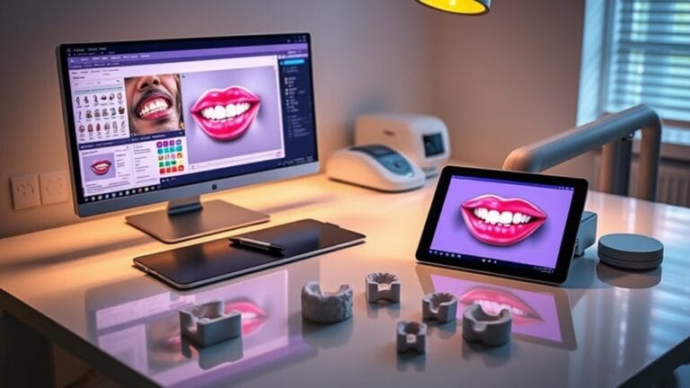
Enhancing Patient Experience With 3D Scans
Utilizing 3D scans greatly enhances the patient experience in orthodontic practices. By eliminating traditional putty molds, you reduce discomfort, making the scanning process quick and non-invasive. Patients only need to keep their mouths open for a few minutes, ensuring no irritation to their lips, gums, or cheeks. This accessibility not only improves comfort but also fosters greater patient engagement.
With 3D technology, you can provide high-quality images that reveal intricate details of dental anatomy, allowing for more accurate diagnoses. Patients appreciate seeing their digital impressions and treatment plans, which helps them understand their oral health better. They can visualize treatment outcomes through simulations, leading to informed decision-making. Additionally, this technology detects hidden oral health issues, which can be crucial for comprehensive care.
Moreover, the ability to view scans instantly streamlines the diagnostic process and reduces the number of visits required. With enhanced communication between you and your patients, their feedback becomes invaluable in tailoring treatment plans. This engagement builds trust, making patients feel more involved and invested in their care.
Frequently Asked Questions
How Does CBCT Differ From Traditional 2D Imaging Methods?
CBCT offers significant advantages over traditional 2D imaging methods, including detailed 3D visualization and reduced radiation exposure. In contrast, 2D limitations often obscure critical anatomical details and can miss early signs of dental issues.
Are 3D Scans Safe for Children and Pregnant Patients?
When traversing the domain of pediatric considerations, 3D scan safety shines bright. These scans offer low radiation and comfort for children and pregnant patients, ensuring accurate diagnoses without compromising health. You're making a wise choice.
How Long Does a Typical 3D Scan Take?
A typical 3D scan duration ranges from 3 to 10 minutes, depending on factors like complexity and resolution. Proper patient preparation guarantees comfort, allowing you to efficiently capture high-resolution images during the process.
What Are the Costs Associated With 3D Imaging Services?
Steering 3D imaging costs is like walking a tightrope; you need balance. Consider the cost comparison between systems and check insurance coverage, as these factors greatly impact your practice's budget and patient accessibility.
Can 3D Imaging Be Used for Dental Emergencies?
Yes, 3D imaging offers significant diagnostic advantages in dental emergencies, enabling accurate treatment planning. It identifies hidden issues quickly, facilitating immediate interventions that enhance patient outcomes and streamline emergency care processes in your practice.
Conclusion
As you explore the future of orthodontics, consider how these cutting-edge 3D imaging solutions can revolutionize your practice. The precision and clarity they offer not only enhance diagnostic capabilities but also pave the way for tailored treatment plans that truly reflect each patient's needs. Imagine the possibilities—improved outcomes, increased patient satisfaction, and a competitive edge in the evolving landscape of dental care.
If you're feeling overwhelmed by the changes or unsure about how to implement these technologies effectively, don't hesitate to reach out to us for assistance. Our expert team can help you navigate these innovations, saving you valuable time, reducing stress, and ultimately improving your dental practice. Are you ready to embrace this transformation and elevate your orthodontic practice to new heights? Let us support you on your journey!