
What Advances Are Revolutionizing Digital X-Ray Technology?
As you explore the domain of digital X-ray technology, you'll notice some remarkable advancements that are reshaping the landscape. Enhanced image quality paired with reduced radiation exposure stands out as a significant shift, driven by innovations like advanced detector systems and AI integration. These developments not only improve diagnostic efficiency but also prioritize patient safety. However, the impact of real-time imaging and 3D capabilities opens up even more questions about the future of this technology. What might these innovations mean for patient care and diagnostics moving forward?
If you have questions or need assistance with this topic, feel free to contact us. We can connect you with experts who can provide valuable solutions and insights.
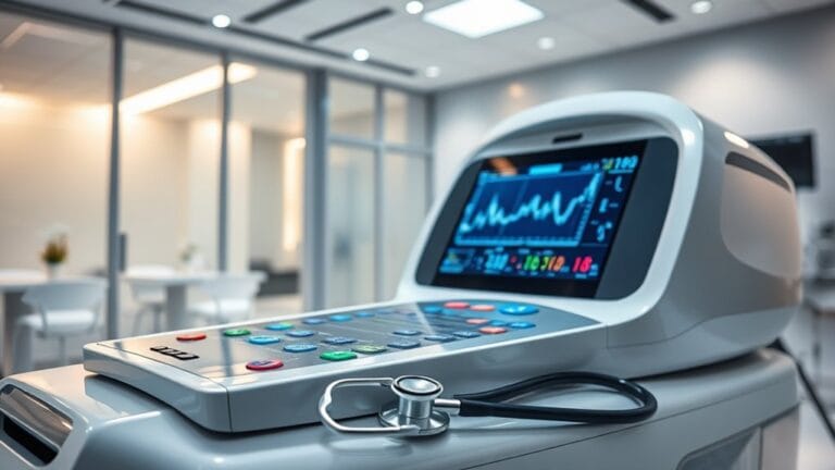
Enhanced Image Quality
As digital X-ray technology evolves, you'll notice significant enhancements in image quality due to advanced detector systems. Newer detectors capture higher resolution images, allowing for finer details in anatomical structures. This improvement directly boosts diagnostic accuracy, enabling you to make more informed decisions regarding patient care.
Advanced materials such as amorphous silicon and amorphous selenium play an essential role in enhancing image resolution and signal-to-noise ratio. Additionally, the expansion of dynamic range facilitates clear visualization of both soft tissue and bony structures within a single image. Moreover, the development of techniques that minimize patient radiation exposure is crucial for ensuring long-term safety during imaging procedures.
Flat-panel detectors contribute to improved image uniformity and faster acquisition times, which streamlines the diagnostic process.
Furthermore, digital images can be easily manipulated for ideal visualization. You can adjust image contrast and brightness, zoom in for closer inspection, and employ AI-powered image processing to enhance quality and precision.
Automated image analysis accelerates diagnosis, ensuring that you can respond quickly to patient needs.
With these advancements, the quality of digital X-rays not only improves but also reinforces your commitment to delivering exceptional care through precise diagnostics.
Reduced Radiation Exposure
Digital X-ray technology greatly reduces radiation exposure, making it a safer option for patients. Compared to conventional X-ray machines, digital systems expose patients to up to 90% less radiation. This reduction is especially important in pediatric imaging, where minimizing radiation is essential for young patients whose developing bodies are more sensitive to cumulative exposure.
Modern digital X-ray systems leverage advanced, more sensitive detectors that capture high-quality images with greatly reduced radiation. Sophisticated software algorithms optimize the balance between image quality and safety, ensuring that patients receive the necessary diagnostic information with minimal risk. Additionally, studies by the American College of Radiology indicate that even low levels of radiation exposure can accumulate over time, reinforcing the importance of using digital X-rays.
In cases where patients need frequent imaging, the cumulative radiation doses over time are reduced, thereby lessening long-term health concerns. Moreover, digital X-rays eliminate the need for repeat exposures, further enhancing patient safety.
The integration of Artificial Intelligence in image analysis helps identify subtle patterns without increasing radiation doses. As awareness of radiation effects grows, the focus on reduced exposure underscores the commitment to patient safety, especially in vulnerable populations like children.
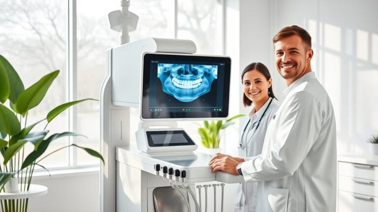
Instant Image Availability
Building on the notable reduction in radiation exposure, digital X-ray technology offers the advantage of instant image availability. This feature grants you immediate access to images almost as soon as they're taken, eliminating the traditional wait times associated with film development. With this capability, you can facilitate quicker assessments and initiate treatment without delay, thereby enhancing patient engagement through instant visual understanding.
Moreover, instant image availability streamlines the diagnostic process, optimizing your workflow considerably. Quick image processing allows for faster, more accurate diagnoses, reducing the necessity for retakes and minimizing additional radiation exposure. This efficiency in medical procedures not only enhances patient care but also supports immediate medical evaluations across various settings. Furthermore, digital images can be easily transferred and enhanced for clarity, improving the overall diagnostic experience.
The clarity and detail of digital images further improve diagnostic accuracy. You can digitally enhance and enlarge these images to analyze internal structures more effectively, detecting minute details and abnormalities at earlier stages.
Additionally, the ease of electronic sharing and storage means you can seamlessly collaborate with other healthcare professionals or insurance companies, further optimizing the workflow and enhancing patient outcomes.
Real-Time Imaging Advances
Real-time imaging advances have revolutionized diagnostic capabilities in modern medicine. By employing cutting-edge image processing algorithms, you can now achieve enhanced speed and accuracy, resulting in clearer, more detailed visualizations of internal structures during procedures. This dynamic imaging technology supports a wide range of clinical applications, greatly improving diagnostic efficiency.
The latest fluoroscopy units incorporate real-time image enhancement, which boosts contrast and detail, thereby improving the visibility of anatomical structures. This enhancement not only augments diagnostic accuracy but also streamlines the process, reducing the need for repeat scans due to superior image quality. As a result, you can make more informed decisions in medical contexts.
Moreover, automated image analysis powered by AI algorithms facilitates rapid interpretation of images, identifying subtle patterns and anomalies that might escape human detection. This capability analyzes vast amounts of data in seconds, speeding up diagnostics while prioritizing critical cases for radiologists. Additionally, advancements in digital radiography technology have further contributed to the overall improvements in imaging quality.
In addition, advanced detector technologies, including high-resolution flat-panel detectors, contribute to improved image clarity and reduced radiation exposure. Collectively, these real-time imaging advances transform the landscape of diagnostics, ultimately enhancing patient outcomes and care quality.
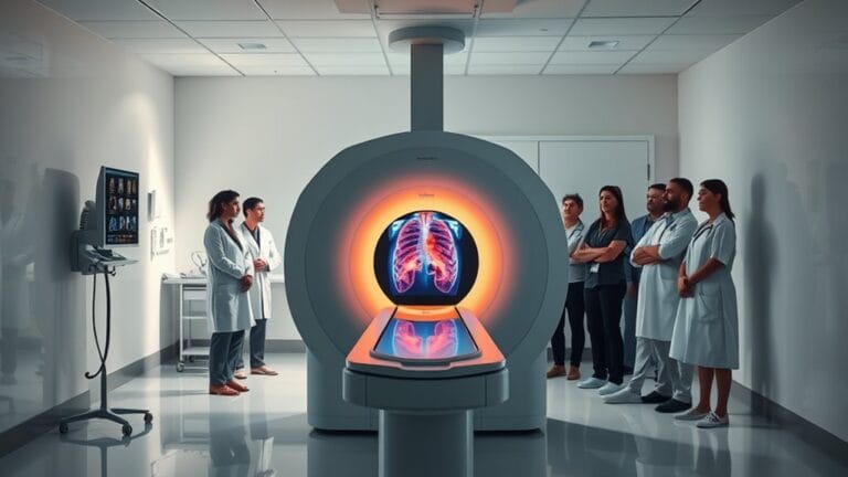
3D Imaging Innovations
Among the latest advancements in medical imaging, D imaging innovations stand out for their ability to enhance diagnostic capabilities considerably.
3D X-ray imaging, for instance, provides a depth and detail that traditional 2D X-rays simply can't match, aiding in the identification of complex issues and improving surgical planning. These advancements allow you to make more informed decisions in patient care.
Here are three key benefits of D imaging innovations:
- Enhanced Diagnostic Accuracy: 3D visualization techniques enable a thorough view of internal structures, ensuring accurate diagnoses.
- Improved Bone Health Assessment: DEXA advancements allow for precise measurement of bone mineral density, facilitating early detection of conditions like osteoporosis.
- AI Integration: By highlighting areas of concern, AI enhances image interpretation, ensuring nothing is overlooked in the diagnostic process. Moreover, AI algorithms can analyze imaging data at high speeds from extensive medical databases to further streamline the diagnostic process.
Together, these innovations are transforming how you approach diagnostics, ultimately leading to better patient outcomes.
With enhanced detector technology, it's possible to obtain clearer images with lower radiation doses, further prioritizing patient safety.
Embracing these advancements positions you to provide exceptional care while ensuring precision in your evaluations.
Portable Imaging Solutions
Advancements in imaging technology have paved the way for portable imaging solutions that greatly enhance patient care. These mobile diagnostics options feature lightweight and compact designs, allowing for easy transportability within healthcare settings. They're specifically engineered for bedside imaging, making them invaluable for patients with limited mobility or those in intensive care.
The shift from traditional film-based X-rays to digital radiography in portable units greatly improves the imaging process. You benefit from immediate image capture and manipulation, leading to higher image quality and reduced radiation exposure. Furthermore, the digital format allows for electronic storage and seamless transmission of images, enhancing diagnostic accuracy. Additionally, less radiation exposure is a key advantage of digital radiography, ensuring safer imaging for all patients.
Wireless capabilities further revolutionize portable imaging. They eliminate cumbersome cables, facilitating real-time image sharing and ensuring efficient integration with hospital networks. This connectivity enhances collaboration among healthcare providers, allowing for quicker diagnoses and treatment plans.
Ultimately, portable imaging solutions reduce patient waiting times and make imaging accessible in remote or challenging settings. By improving overall efficiency in patient care, these advancements demonstrate a commitment to serving patients effectively and compassionately.
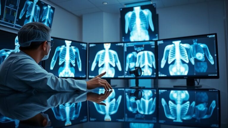
AI in Image Interpretation
Harnessing the power of artificial intelligence (AI) in image interpretation is revolutionizing diagnostic radiology. With advancements in AI diagnostics, you can expect significant improvements in both accuracy and efficiency. AI algorithms now perform at levels comparable to or exceeding those of human radiologists, detecting subtle abnormalities that might be missed by the human eye. This transformation emphasizes three key benefits:
- Improving Diagnostic Accuracy: AI enhances precision, particularly in complex cases like cancer and neurological disorders, reducing human error.
- Streamlining Workflow: By automating mundane tasks, AI accelerates image processing and analysis, allowing you to handle high case volumes without fatigue.
- Advanced AI Models: Transformer-based models generate radiology reports akin to those produced by radiologists, maintaining clinical accuracy and textual quality. Moreover, AI algorithms demonstrate performance comparable to human radiologists, highlighting their potential to enhance diagnostic practices.
The integration of AI with electronic health records (EHRs) and its potential in personalized medicine mark a significant shift in how we approach medical imaging.
As you embrace these innovations, ongoing research will be essential to validate AI's reliability and efficacy, ultimately transforming the radiography industry for the better.
Frequently Asked Questions
How Do Advancements Improve Patient Comfort During X-Ray Procedures?
Advancements in digital X-ray technology enhance your patient experience by providing improved imaging quality with thinner sensors. This results in less discomfort, faster procedures, and an overall more pleasant experience during necessary diagnostic evaluations.
What Are the Cost Implications of Transitioning to Digital X-Ray Systems?
Shifting to digital X-ray systems is like investing in a high-performance vehicle; while the initial investment is substantial, the long-term savings through reduced operational costs and improved efficiency can greatly enhance your practice's financial health.
How Is Data Security Handled in Digital Radiography?
In digital radiography, you handle data security through robust data encryption, strict access controls, and compliance standards. Regular software updates and secure image storage safeguard patient privacy, ensuring sensitive information remains protected from unauthorized access.
What Training Is Required for Radiologists Using New Technologies?
To effectively use new technologies, you need thorough training that combines virtual training and hands-on experience. This approach guarantees you grasp digital radiography principles and advanced techniques, optimizing your skills for superior patient care and diagnostics.
How Do These Advancements Impact Patient Throughput in Clinics?
These advancements enhance patient throughput by streamlining workflow optimization and improving patient scheduling. Faster imaging and processing reduce wait times, allowing you to serve more patients effectively while ensuring quality diagnostic care.
Conclusion
As you navigate the evolving landscape of digital X-ray technology, imagine a world where crystal-clear images emerge instantly, revealing intricate details with minimal radiation. Picture yourself in a clinic, where AI swiftly interprets scans, guiding healthcare professionals to accurate diagnoses. With portable devices and real-time imaging at your fingertips, patient care transforms into a seamless experience. These advancements not only enhance diagnostic precision but also prioritize safety, ensuring that every patient receives the best possible care in a timely manner.
If you have questions or need assistance with integrating these revolutionary technologies into your practice, don't hesitate to reach out to us. Our expert help can save you time, reduce stress, and ultimately improve your dental practice. Embrace the future of dental care with confidence, knowing that support is just a call away!